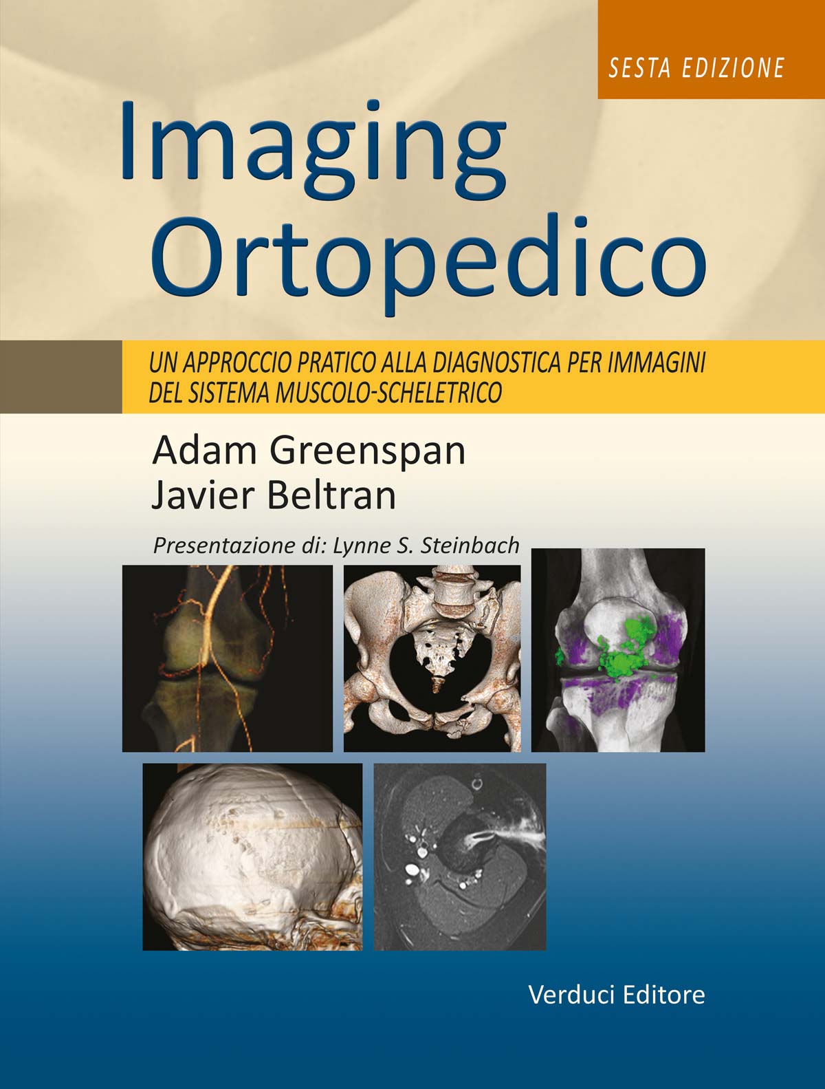
Greenspan A. - Beltran J.
Imaging Ortopedico
LAST COPIES AVAILABLE
Large-format volume (30.5 × 23.5 cm), 1184 pages, fully illustrated in colour.
The previous edition of this work, Orthopaedic Radiology, established itself as a comprehensive reference and an ideal guide to musculoskeletal imaging for radiologists and orthopaedic surgeons at all levels of experience.
This sixth edition, Imaging Ortopedico – A Practical Approach to Musculoskeletal Diagnostic Imaging, reaffirms its founding principles while expanding its objectives, enabling readers to interpret a wide range of findings supported by more than 4,000 high-quality images.
-
Effectively interpret findings obtained through conventional radiography, ultrasound,
computed tomography (CT), dual-energy CT, PET-CT, magnetic resonance imaging (MRI)
and three-dimensional (3D) imaging. -
Gain mastery of current trends in orthopaedic radiology, with increasing emphasis on
ultrasound and MRI compared with higher-radiation techniques. -
Select the most appropriate imaging approach for each patient, considering accuracy,
speed and cost. -
Apply up-to-date knowledge in MRI interpretation guided by leading authorities in
musculoskeletal imaging. -
Quickly access essential information through diagrams, summary tables and “Practical
Points to Remember” sections at the end of each chapter.
€0,00
Out of stock
Additional information
| edizione | LUGLIO 2016 |
|---|---|
| autori | Greenspan A., Beltran J. |
| formato | 30,5 x 23,5 |
| pagine | 1184 pagine |
| informazioni extra | interamente a colori |
Description
PART I – Introduction to diagnostic imaging in orthopaedics
- Chapter 1 – Role of the orthopaedic radiologist
- Chapter 2 – Diagnostic imaging techniques in orthopaedics
- Chapter 3 – Bone formation and growth
PART II – Trauma
- Chapter 4 – Radiological assessment of trauma
- Chapter 5 – Upper limb I – Shoulder girdle
- Chapter 6 – Upper limb II – Elbow
- Chapter 7 – Upper limb III – Distal forearm, wrist and hand
- Chapter 8 – Lower limb I – Pelvic girdle, sacrum and proximal femur
- Chapter 9 – Lower limb II – Knee
- Chapter 10 – Lower limb III – Ankle and foot
- Chapter 11 – Spine
PART III – Arthritis
- Chapter 12 – Radiological evaluation of arthritis
- Chapter 13 – Joint disease
- Chapter 14 – Inflammatory arthritis
- Chapter 15 – Other arthritides and arthropathies
PART IV – Tumours and pseudotumoural lesions
- Chapter 16 – Radiological evaluation of tumours and pseudotumoural lesions
- Chapter 17 – Benign tumours and pseudotumours I – Osteogenic lesions
- Chapter 18 – Benign tumours and pseudotumours II – Cartilaginous lesions
- Chapter 19 – Benign tumours and pseudotumours III – Fibrous, osteophytic and fibrohistiocytic lesions
- Chapter 20 – Benign tumours and pseudotumours IV – Other benign lesions
- Chapter 21 – Malignant bone tumours I – Osteosarcomas and chondrosarcomas
- Chapter 22 – Malignant bone tumours II – Other tumours
- Chapter 23 – Tumours and pseudotumours of the joints
PART V – Infections
- Chapter 24 – Radiological evaluation of musculoskeletal infections
- Chapter 25 – Osteomyelitis, septic arthritis and soft tissue infections
PART VI – Metabolic and endocrine disorders
- Chapter 26 – Radiological evaluation of metabolic and endocrine disorders
- Chapter 27 – Osteoporosis, rickets and osteomalacia
- Chapter 28 – Hyperparathyroidism
- Chapter 29 – Paget’s disease
- Chapter 30 – Other metabolic and endocrine disorders
PART VII – Congenital and developmental abnormalities
- Chapter 31 – Radiological evaluation of skeletal malformations
- Chapter 32 – Upper and lower limb malformations
- Chapter 33 – Scoliosis and generalized skeletal abnormalities
Related products
-
 Andrews J.R. - Harrelson G.L. - Wilk K.E.
Riabilitazione nella Traumatologia dello Sport
Original price was: €100,00.€95,00Current price is: €95,00. Buy now -
 Brinker M.R. - Miller M.D.
Principi e Pratica di Ortopedia
Original price was: €48,00.€45,60Current price is: €45,60. Buy now -
Romanini L.
La Bella Storia di Raffaele Paparella-Treccia
Original price was: €15,00.€14,25Current price is: €14,25. Buy now