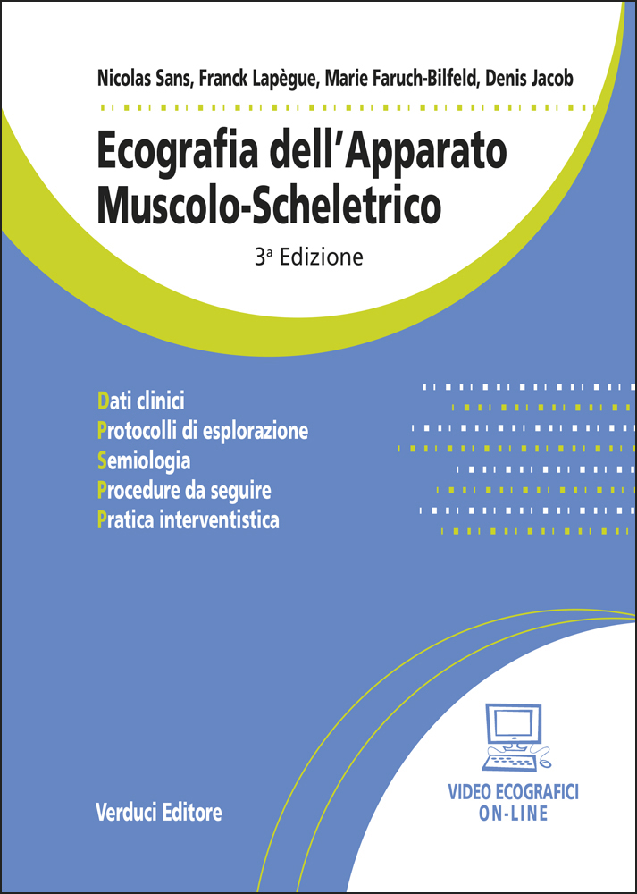
Ecografia dell’Apparato Muscolo-Scheletrico
14 x 21 cm format, 404 pages, approximately 500 colour images and numerous tables, hardcover laminated colour binding. Online ultrasound videos included.
3rd Edition.
Ultrasound is the first-line examination for tendon, joint and muscle injuries, whether traumatic or not. Learning this technique is delicate but essential and requires constant updating.
This book serves as a practical reference in the field. It is structured as a hands-on training manual organised by anatomical structures and regions: muscles, tendons, nerves and plexuses, shoulder, elbow, wrist, fingers, hip, knee, ankle and tarsus, and foot. Each chapter first outlines the normal appearance and anatomical characteristics of a structure or joint, an indispensable prerequisite, before addressing pathological conditions starting from clinical symptoms.
This extensively updated edition has been enriched with contemporary iconography. To strengthen its pedagogical value, the authors provide clear and precise sono-anatomical diagrams and place ultrasound in perspective with other imaging modalities within a rational patient-oriented framework.
Online videos accompany the book. They illustrate the most frequently performed interventional procedures, including approach techniques, environmental conditions and precautions to be taken before any invasive procedure.
The book is intended for radiologists, sports medicine physicians, rheumatologists and orthopaedic surgeons.
Original price was: €70,00.€66,50Current price is: €66,50.
Additional information
| edizione | 3 edizione Marzo 2023 |
|---|---|
| autori | Sans N., Lapegue F., Faruch-Bilfeld M., Jacob D. |
| formato | Volume formato 14 x 21 |
| pagine | 404 pagine |
| informazioni extra | numerose tabelle, circa 500 immagini a colori, Video Ecografici on-line, rilegato con copertina a colori e plastificata |
Description
Table of Contents
Chapter 1 – Tendons
- Normal tendon
- Pathological tendon
Chapter 2 – Muscles
- Normal muscle and physiological variants
- Pathological muscle
Chapter 3 – Nerves and Plexuses
- Normal nerve
- Pathological nerve
- Normal brachial plexus
- Pathological brachial plexus
Chapter 4 – Shoulder
- Normal shoulder
- Pathological shoulder
Chapter 5 – Elbow
- Normal elbow
- Pathological elbow
Chapter 6 – Wrist
- Normal wrist
- Pathological wrist
Chapter 7 – Fingers
- Normal fingers
- Finger disorders
Chapter 8 – Hip
- Normal hip
- Pathological hip
Chapter 9 – Knee
- Normal knee
- Pathological knee
- Prosthetic knee
Chapter 10 – Ankle and tarsus (excluding forefoot)
- Normal ankle
- Pathological ankle
Chapter 11 – Foot
- Normal foot
- Pathological foot
Chapter 12 – Interventional ultrasound
- General considerations
- Most frequently performed procedures
Index
Online supplements
Online videos are available covering the most frequently performed procedures.
- Video 1 – Subacromial-subdeltoid bursa injection
- Video 2 – Bicipital recess and glenohumeral joint injection
- Video 3 – Acromioclavicular joint injection
- Video 4 – Injection for lateral epicondylitis
- Video 5 – Injection for De Quervain’s tenosynovitis
- Video 6 – Carpal tunnel injection
- Video 7 – Trigger finger injection
- Video 8 – Peritrochanteric injection
- Video 9 – Psoas impingement injection
- Video 10 – Musculocutaneous nerve injection
- Video 11 – Aspiration and injection of a popliteal cyst
- Video 12 – Iliotibial band injection
- Video 13 – Evacuative puncture of a muscular haematoma
- Video 14 – Superficial plantar fascia injection
- Video 15 – Posterior tibial tendon tenosynovitis injection
- Video 16 – Intermetatarsal injection for Morton’s neuroma
Related products
-
 Fernando Alemanno
Invecchiare bene Vivere meglio
Original price was: €15,00.€10,90Current price is: €10,90. Buy now -
 Alexander N. Sencha, Yury N. Patrunov
Ecografia Tiroidea
Original price was: €68,00.€64,50Current price is: €64,50. Buy now -

Diagnosi e Trattamento delle Lesioni Traumatiche dei Nervi Periferici – Raccomandazioni della Società Italiana di Microchirurgia
Original price was: €60,00.€57,00Current price is: €57,00. Buy now