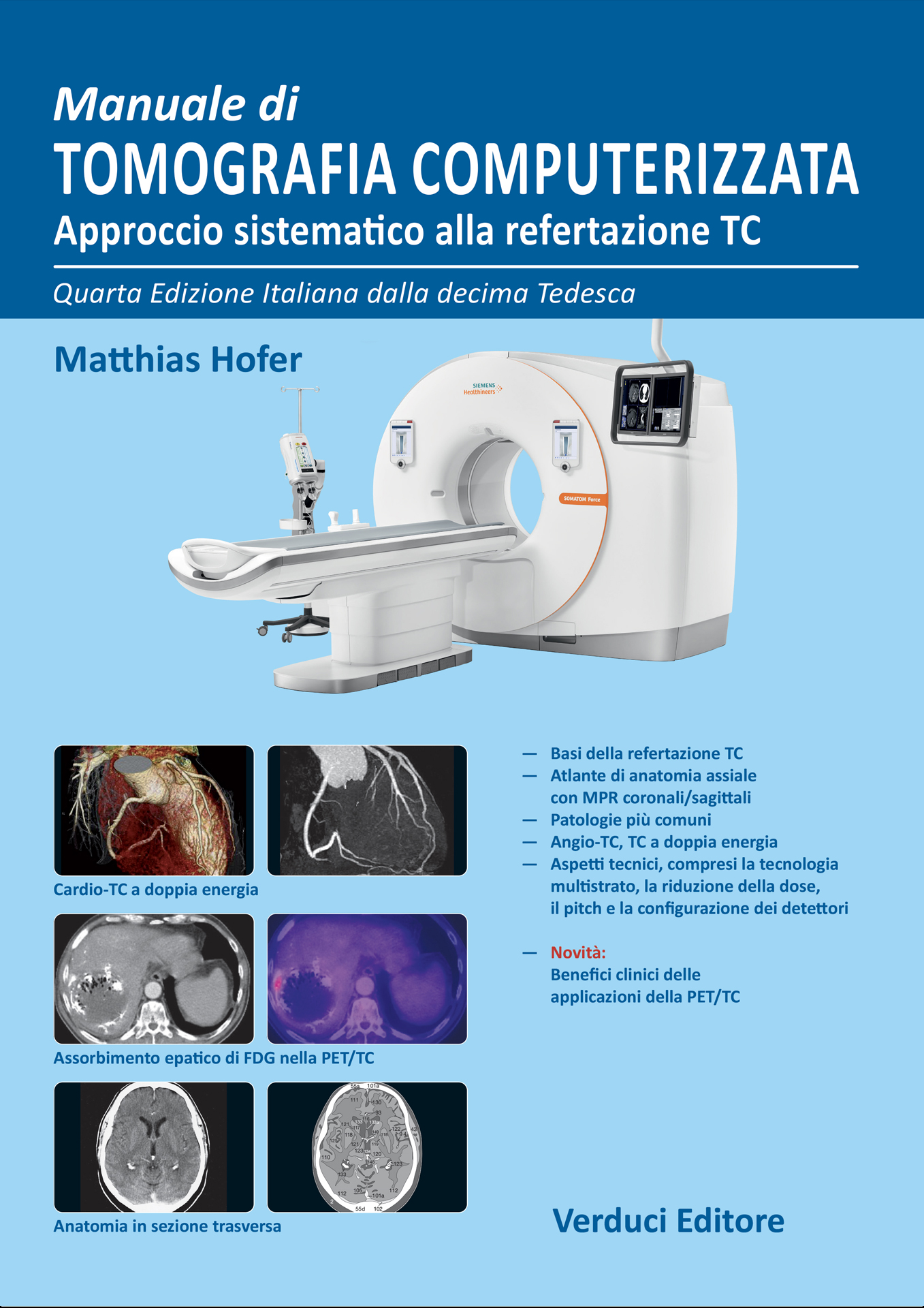
Manuale di Tomografia Computerizzata Approccio sistematico alla refertazione TC
4th Italian Edition from the 10th German Edition
Large-format volume (22 x 30 cm), 232 pages with 1300 images, printed in full color on matte coated paper and bound in paperback with laminated color cover.
Computed tomography (CT) has become an integral and indispensable part of clinical diagnostics, allowing highly accurate answers to specific clinical questions within a short time frame. Recent technological advances enable submillimeter spatial resolution in CT angiography and, with the aid of dual-energy CT, even characterization of the chemical composition of tissues or, for example, renal calculi.
Above all, the development of PET/CT has made possible tremendous progress in oncology, particularly regarding diagnostic accuracy in the detection of metastases and tumor recurrence.
Modern imaging modalities are often not taught adequately during clinical lectures and conferences for medical students, who frequently graduate with significant knowledge gaps in this field.
All these aspects and recent advances have already been taken into account in this standard manual of CT diagnostics, which provides the necessary basic knowledge for beginners who are just becoming familiar with the subject, while also serving advanced users with a special radiological interest.
It is hoped that the quizzes, in particular, will stimulate many readers to test and refine their diagnostic skills.
The success story of 28 editions in 10 languages speaks for itself and demonstrates the wide acceptance of this book among German-speaking colleagues as well as international professionals.
I wish readers great benefit, both in terms of learning and enjoyment, from using this educational manual.
Original price was: €85,00.€80,50Current price is: €80,50.
Additional information
| edizione | 2024 (4 Edizione) |
|---|---|
| autori | Hofer M. |
| formato | Volume grande formato (22 x 30) |
| pagine | 232 pagine |
| informazioni extra | stampato a colori su carta patinata opaca e rilegato in brossura con copertina stampata a colori e plastificata. |
Description
- Fundamentals of CT reporting
- Atlas of axial anatomy with coronal and sagittal MPR reconstructions
- Most common pathologies
- CT Angiography and Dual-Energy CT
- Technical aspects: multislice technology, dose reduction, pitch and detector configuration
- New: Clinical benefits of PET/CT applications
Contents
- Physical and Technical Principles
- Principles of CT Image Interpretation
- Patient Preparation
- Administration of Contrast Media
- CT of the Brain
- CT of the Brain: Normal Findings
- Brain Pathology
- Cervical CT
- Cervical Pathology
- CT of the Chest
- CT of the Chest: Normal and Pathological Findings
- CT of the Abdomen
- CT of the Abdomen: Pathological Findings
- Spine and Skeletal Pathology
- Lower Limb
- Interventional CT
- The “ABC” of CT Interpretation
- Radiation Protection
- CT Angiography
- Contrast Media Injectors
- Dual-Energy CT: Technique
- Dual-Energy CT: Applications
- PET/CT: Introduction
- Anatomy in Coronal MPR
- Anatomy in Sagittal MPR
Related products
-
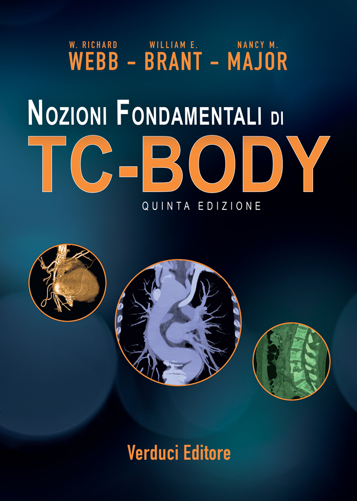
 WEBB W.R. - BRANT W.E. - MAJOR N.M.
Nozioni Fondamentali di TC-BODY – 5th Edition
Original price was: €95,00.€90,00Current price is: €90,00. Buy now -
 Potestà P.
Manuale-Prontuario di Diagnostica e Terapia Medica
Original price was: €49,00.€25,00Current price is: €25,00. Buy now