Sale!
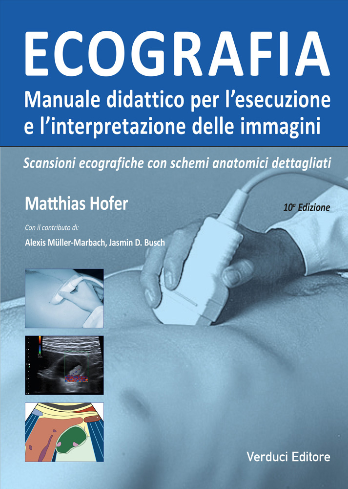
Mathias Hofer
Ecografia Manuale didattico
Ultrasound – Educational Manual.
The text is structured in lessons and guides you like a tutor through a systematic course for the examination of individual organs and systems.
- Numerous images clearly illustrate the correct probe positioning.
- Triple representation: probe position, ultrasound image, and schematic drawing of topographic anatomy.
- The numerical coding shown in the schematic drawings allows you to stimulate your learning and verify it at any time.
- The numerical summary system is valid for every page of the text.
- The quiz images at the end of each chapter allow you to assess your learning progress in an engaging and interactive way.
- The principles of physics are not presented in a sterile manner, but through easy-to-understand diagrams and illustrations.
- Numerous practical tips facilitate the approach to this methodology.
- The practical “ABC” of ultrasound findings provides useful terminology.
Original price was: €61,00.€58,00Current price is: €58,00.
Additional information
| formato | 21,5 x 30 |
|---|---|
| pagine | 166 pagine |
| informazioni extra | stampato a colori rilegato in brossura copertina a colori e plastificata. Numerose Illustrazioni schemi e tabelle. |
| Autori | Mathias Hofer |
Description
Introduction
- Principles of physics/technique
- Innovative methodologies
- Artifacts
- Self-assessment quiz
- Practical tips for beginners
Lesson 1 – Retroperitoneum, sagittal plane
- Anatomy
- Normal findings
- Aortic aneurysm
- Right heart failure
- Self-assessment quiz
Lesson 2 – Retroperitoneum, transverse plane
- Anatomy
- Normal findings
- Pancreatitis
- Pancreatic tumors
- Retroperitoneal lymph nodes
- Self-assessment quiz
Lesson 3 – Hepatic hilum, gallbladder, bile ducts
- Anatomy
- Hepatic hilum:
- Normal findings
- Portal hypertension
- Lymph nodes, portal vein thrombosis
- Gallbladder:
- Cholecystitis
- Gallstones
- Gallbladder polyps
- Cholestasis
- Self-assessment quiz (after Lesson 4)
Lesson 4 – Liver
- Anatomy of hepatic segments
- Normal findings: organ size, margins, angles
- Confluence of hepatic veins, right heart failure
- Anatomical variants, hepatic steatosis
- Hepatic cysts
- Hepatic hemangiomas
- Focal nodular hyperplasia (FNH)
- Liver cirrhosis
- Hepatocellular carcinoma (HCC)
- Liver metastases
- Self-assessment quiz (Lessons 3 and 4)
Lesson 5 – Adrenal glands, kidneys, transplanted kidney and spleen
- Kidneys and adrenal glands:
- Anatomy
- Normal findings
- Anatomical variants and renal cysts
- Renal degeneration and nephritis
- Urinary stasis/obstruction
- Differential diagnosis of urinary stasis
- Renal stones and infarctions
- Renal and adrenal tumors
- Transplanted kidney
- Spleen:
- Anatomy and examination technique
- Spleen dimensions
- Splenomegaly and splenic infarction
- Focal splenic lesions
- Self-assessment quiz
Lesson 6 – Thyroid gland, lymph nodes, gastrointestinal tract (GIT)
- Thyroid:
- Anatomy and volumetry
- Normal findings
- Goiter
- Thyroid nodules, thyroiditis
- Lymph nodes (LN):
- Lateral cervical lymph nodes
- Differential diagnostic criteria, perfusion parameters
- Differential diagnostic criteria, metastatic LN
- Retroperitoneal LN
- Gastrointestinal tract (GIT):
- Wall anatomy of the GIT
- Gastric neoplasms
- Crohn’s disease, fistulas
- Intestinal intussusception, hernias
- Sprue, diarrhea, appendicitis
- Colon
- Diverticulitis
- Self-assessment quiz
Lesson 7 – Bladder and genital organs
- Bladder:
- Anatomy
- Normal findings
- Indwelling catheter and differential diagnosis of cystitis
- Genital organs:
- Prostate and testicles
- Undescended testis, orchitis, hydrocele
- Female genital organs
- Uterus: normal findings
- Uterine tumors
- Ovaries: normal findings
- Ovarian cysts and tumors
- Diagnosis of pregnancy
- Placenta and sex determination
- Self-assessment quiz
Lesson 8 – FAST, eFAST and lung
- FAST algorithms
- eFAST algorithms
- Seashore sign, Barcode sign
- Pleural sliding, lung pulse in apnea
- Lung point in pneumothorax
- Pleura:
- Quantification of pleural effusion
- Pleuritis, empyema, mesothelioma
- Ribs:
- Rib fractures, rib metastases
- Lung:
- Pneumonia, infarction, bronchial carcinoma
- Self-assessment quiz
Lesson 9 – Pediatrics: Skull/CNS, hip joint, kidneys
- Skull/CNS:
- Anatomy of cerebrospinal fluid spaces
- Sagittal scan: normal findings
- Anatomical variants
- Coronal scan: normal findings
- Cerebral hemorrhages
- Hydrocephalus
- Spinal canal
- Hip joint:
- Preparation and positioning
- Normal findings
- Calibration and measurement errors
- Graf classification
- Kidneys and urinary tract:
- Neonatal kidney
- Stasis and reflux
- Renal and adrenal tumors
- Urachus, ureterocele, spleen dimensions
- GIT:
- Pyloric hypertrophy, reflux, Hirschsprung’s disease
Related products
-
 Major - Anderson - Helms - Kaplan - Dussault
RM Muscolo-Scheletrica
Original price was: €150,00.€142,50Current price is: €142,50. Buy now -
 Potestà P.
Manuale-Prontuario di Diagnostica e Terapia Medica
Original price was: €49,00.€25,00Current price is: €25,00. Buy now -

Diagnosi e Trattamento delle Lesioni Traumatiche dei Nervi Periferici – Raccomandazioni della Società Italiana di Microchirurgia
Original price was: €60,00.€57,00Current price is: €57,00. Buy now